

Another Name For An Elbow

A hand drawn prototype of the within of an elbow joint with annotations in an up-close way
| Elbow | |
|---|---|
 | |
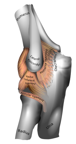 Beefcake of the elbow (left). | |
| Details | |
| Identifiers | |
| Latin | articulatio cubiti |
| MeSH | D004550 |
| TA2 | 145 |
| FMA | 24901 |
| Anatomical terminology [edit on Wikidata] | |
The elbow is the region betwixt the arm and the forearm that surrounds the elbow joint.[ane] The elbow includes prominent landmarks such equally the olecranon, the cubital fossa (too called the chelidon, or the elbow pit), and the lateral and the medial epicondyles of the humerus. The elbow joint is a swivel joint between the arm and the forearm;[two] more specifically between the humerus in the upper arm and the radius and ulna in the forearm which allows the forearm and mitt to be moved towards and away from the trunk.[3] [4] The term elbow is specifically used for humans and other primates, and in other vertebrates forelimb plus joint is used.[i]
The name for the elbow in Latin is cubitus, so the word cubital is used in some elbow-related terms, as in cubital nodes for example.
Construction [edit]
Joint [edit]
The elbow joint has three unlike portions surrounded by a common articulation capsule. These are joints betwixt the 3 bones of the elbow, the humerus of the upper arm, and the radius and the ulna of the forearm.
| Joint | From | To | Description |
|---|---|---|---|
| Humeroulnar joint | trochlear notch of the ulna | trochlea of humerus | Is a elementary hinge-joint, and allows for movements of flexion and extension just. |
| Humeroradial joint | head of the radius | capitulum of the humerus | Is a ball-and-socket articulation. |
| Proximal radioulnar articulation | caput of the radius | radial notch of the ulna | In any position of flexion or extension, the radius, carrying the hand with it, tin be rotated in it. This motion includes pronation and supination. |
When in anatomical position there are four principal bony landmarks of the elbow. At the lower office of the humerus are the medial and lateral epicondyles, on the side closest to the body (medial) and on the side away from the body (lateral) surfaces. The tertiary landmark is the olecranon found at the head of the ulna. These lie on a horizontal line called the Hueter line. When the elbow is flexed, they form a triangle called the Hueter triangle, which resembles an equilateral triangle.[5]
At the surface of the humerus where it faces the joint is the trochlea. In most people, the groove running across the trochlea is vertical on the anterior side just it spirals off on the posterior side. This results in the forearm being aligned to the upper arm during flexion, but forming an angle to the upper arm during extension — an angle known as the carrying angle.[6]
The superior radioulnar joint shares the joint capsule with the elbow articulation but plays no functional role at the elbow.[7]
Joint capsule [edit]

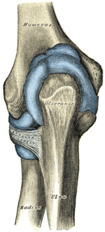
Capsule of elbow-joint (distended). Anterior and posterior aspects.
The elbow joint and the superior radioulnar joint are enclosed past a single fibrous capsule. The capsule is strengthened by ligaments at the sides merely is relatively weak in front and behind.[8]
On the anterior side, the sheathing consists mainly of longitudinal fibres. However, some bundles amid these fibers run obliquely or transversely, thickening and strengthening the capsule. These bundles are referred to every bit the capsular ligament. Deep fibres of the brachialis musculus insert anteriorly into the sheathing and human action to pull information technology and the underlying membrane during flexion in order to forbid them from existence pinched.[8]
On the posterior side, the capsule is thin and mainly equanimous of transverse fibres. A few of these fibres stretch across the olecranon fossa without attaching to information technology and form a transverse ring with a free upper border. On the ulnar side, the capsule reaches downward to the posterior function of the annular ligament. The posterior sheathing is attached to the triceps tendon which prevents the capsule from existence pinched during extension.[viii]
Synovial membrane [edit]
The synovial membrane of the elbow joint is very extensive. On the humerus, it extends up from the articular margins and covers the coronoid and radial fossae anteriorly and the olecranon fossa posteriorly. Distally, it is prolonged downwards to the neck of the radius and the superior radioulnar joint. It is supported by the quadrate ligament below the annular ligament where it also forms a fold which gives the head of the radius freedom of movement.[8]
Several synovial folds project into the recesses of the articulation.[8] These folds or plicae are remnants of normal embryonic development and can be categorized every bit either anterior (anterior humeral recess) or posterior (olecranon recess).[9] A crescent-shaped fold is usually present between the head of the radius and the capitulum of the humerus.[8]
On the humerus there are extrasynovial fat pads side by side to the iii articular fossae. These pads make full the radial and coronoid fossa anteriorly during extension, and the olecranon fossa posteriorly during flexion. They are displaced when the fossae are occupied past the bony projections of the ulna and radius.[8]
Ligaments [edit]


Left elbow-articulation
Left: anterior and ulnar collateral ligaments
Right: posterior and radial collateral ligaments
The elbow, like other joints, has ligaments on either side. These are triangular bands which alloy with the joint capsule. They are positioned so that they always lie beyond the transverse joint axis and are, therefore, always relatively tense and impose strict limitations on abduction, adduction, and centric rotation at the elbow.[8]
The ulnar collateral ligament has its noon on the medial epicondyle. Its inductive ring stretches from the anterior side of the medial epicondyle to the medial edge of the coronoid process, while the posterior ring stretches from posterior side of the medial epicondyle to the medial side of the olecranon. These two bands are separated past a thinner intermediate function and their distal attachments are united by a transverse ring below which the synovial membrane protrudes during joint movements. The anterior ring is closely associated with the tendon of the superficial flexor muscles of the forearm, even being the origin of flexor digitorum superficialis. The ulnar nervus crosses the intermediate part equally it enters the forearm.[8]
The radial collateral ligament is attached to the lateral epicondyle beneath the mutual extensor tendon. Less singled-out than the ulnar collateral ligament, this ligament blends with the annular ligament of the radius and its margins are attached near the radial notch of the ulna.[8]
Muscles [edit]
Flexion [edit]
There are 3 main flexor muscles at the elbow:[10]
- Brachialis acts exclusively as an elbow flexor and is one of the few muscles in the man body with a single function. It originates low on the anterior side of the humerus and is inserted into the tuberosity of the ulna.
- Brachioradialis acts essentially as an elbow flexor but also supinates during extreme pronation and pronates during farthermost supination. Information technology originates at the lateral supracondylar ridge distally on the humerus and is inserted distally on the radius at the styloid process.
- Biceps brachii is the main elbow flexor but, every bit a biarticular muscle, also plays important secondary roles as a stabiliser at the shoulder and as a supinator. It originates on the scapula with two tendons: That of the long caput on the supraglenoid tubercle just to a higher place the shoulder joint and that of the short head on the coracoid procedure at the acme of the scapula. Its primary insertion is at the radial tuberosity on the radius.
Brachialis is the main muscle used when the elbow is flexed slowly. During rapid and forceful flexion all three muscles are brought into activeness assisted past the superficial forearm flexors originating at the medial side of the elbow.[11] The efficiency of the flexor muscles increases dramatically equally the elbow is brought into midflexion (flexed 90°) — biceps reaches its angle of maximum efficiency at 80–ninety° and brachialis at 100–110°.[x]
Active flexion is express to 145° by the contact between the anterior muscles of the upper arm and forearm, more so because they are hardened by wrinkle during flexion. Passive flexion (forearm is pushed against the upper arm with flexors relaxed) is express to 160° past the bony projections on the radius and ulna every bit they accomplish to shallow depressions on the humerus; i.east. the caput of radius being pressed against the radial fossa and the coronoid procedure existence pressed confronting the coronoid fossa. Passive flexion is further limited by tension in the posterior capsular ligament and in triceps brachii.[12]
A small accessory muscle, and so chosen epitrochleoanconeus muscle, may be found on the medial attribute of the elbow running from the medial epicondyle to the olecranon.[13]
Extension [edit]
Elbow extension is merely bringing the forearm back to anatomical position.[xi] This activeness is performed by triceps brachii with a negligible assist from anconeus. Triceps originates with two heads posteriorly on the humerus and with its long caput on the scapula but beneath the shoulder joint. Information technology is inserted posteriorly on the olecranon.[10]
Triceps is maximally efficient with the elbow flexed 20–xxx°. Equally the angle of flexion increases, the position of the olecranon approaches the principal axis of the humerus which decreases muscle efficiency. In full flexion, nevertheless, the triceps tendon is "rolled up" on the olecranon equally on a pulley which compensates for the loss of efficiency. Because triceps' long caput is biarticular (acts on two joints), its efficiency is also dependent on the position of the shoulder.[10]
Extension is limited past the olecranon reaching the olecranon fossa, tension in the inductive ligament, and resistance in flexor muscles. Forced extension results in a rupture in one of the limiting structures: olecranon fracture, torn capsule and ligaments, and, though the muscles are normally left unaffected, a bruised brachial avenue.[12]
Blood supply [edit]


The anastomosis and deep veins around the elbow-articulation
The arteries supplying the joint are derived from an extensive circulatory anastomosis between the brachial avenue and its terminal branches. The superior and inferior ulnar collateral branches of the brachial artery and the radial and centre collateral branches of the profunda brachii artery descend from above to reconnect on the joint sheathing, where they as well connect with the anterior and posterior ulnar recurrent branches of the ulnar avenue; the radial recurrent branch of the radial avenue; and the interosseous recurrent branch of the common interosseous artery.[14]
The claret is brought dorsum by vessels from the radial, ulnar, and brachial veins. There are 2 sets of lymphatic nodes at the elbow, normally located above the medial epicondyle — the deep and superficial cubital nodes (likewise called epitrochlear nodes). The lymphatic drainage at the elbow is through the deep nodes at the bifurcation of the brachial artery, the superficial nodes drain the forearm and the ulnar side of the hand. The efferent lymph vessels from the elbow go along to the lateral grouping of axillary lymph nodes.[14] [fifteen]
Nerve supply [edit]
The elbow is innervated anteriorly by branches from the musculocutaneous, median, and radial nerve, and posteriorly from the ulnar nerve and the co-operative of the radial nerve to anconeus.[14]
Development [edit]
The elbow undergoes dynamic development of ossification centers through infancy and adolescence, with the society of both the appearance and fusion of the apophyseal growth centers being crucial in assessment of the pediatric elbow on radiograph, in gild to distinguish a traumatic fracture or apophyseal separation from normal evolution. The social club of appearance can be understood by the mnemonic CRITOE, referring to the capitellum, radial head, internal epicondyle, trochlea, olecranon, and external epicondyle at ages 1, iii, 5, 7, 9 and xi years. These apophyseal centers and so fuse during adolescence, with the internal epicondyle and olecranon fusing last. The ages of fusion are more variable than ossification, but normally occur at xiii, 15, 17, xiii, 16 and 13 years, respectively.[16] In add-on, the presence of a joint effusion can exist inferenced by the presence of the fat pad sign, a structure that is normally physiologically present, but pathologic when elevated by fluid, and always pathologic when posterior.[17]
Function [edit]
The function of the elbow joint is to extend and flex the arm grasp and reach for objects.[18] The range of movement in the elbow is from 0 degrees of elbow extension to 150 degrees of elbow flexion.[19] Muscles contributing to function are all flexion (biceps brachii, brachialis, and brachioradialis) and extension muscles (triceps and anconeus).
In humans, the primary task of the elbow is to properly identify the hand in infinite by shortening and lengthening the upper limb. While the superior radioulnar joint shares joint capsule with the elbow joint, it plays no functional part at the elbow.[vii]
With the elbow extended, the long axis of the humerus and that of the ulna coincide.[twenty] At the same fourth dimension, the articular surfaces on both basic are located in front of those axes and deviate from them at an angle of 45°.[21] Additionally, the forearm muscles that originate at the elbow are grouped at the sides of the articulation in order not to interfere with its movement. The wide angle of flexion at the elbow made possible by this arrangement — almost 180° — allows the basic to be brought nigh in parallel to each other.[7]
Carrying angle [edit]
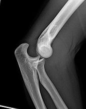
Normal radiograph; correct picture of the straightened arm shows the conveying bending of the elbow
When the arm is extended, with the palm facing forward or up, the bones of the upper arm (humerus) and forearm (radius and ulna) are not perfectly aligned. The divergence from a directly line occurs in the direction of the thumb, and is referred to as the "carrying angle".[22]
The carrying angle permits the arm to exist swung without contacting the hips. Women on average have smaller shoulders and wider hips than men, which tends to produce a larger carrying bending (i.e., larger deviation from a straight line than that in men). There is, all the same, extensive overlap in the carrying angle between individual men and women, and a sexual practice-bias has not been consistently observed in scientific studies.[23]
The angle is greater in the ascendant limb than the non-ascendant limb of both sexes,[24] suggesting that natural forces interim on the elbow modify the carrying angle. Developmental,[25] aging and perchance racial influences add farther to the variability of this parameter.
Pathology [edit]

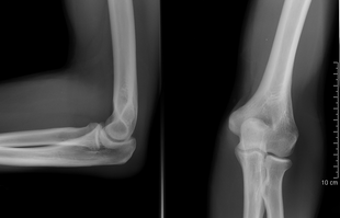
Left: Lateral X ray of a confused right elbow
Right: AP Ten ray of a dislocated correct elbow
The types of disease most commonly seen at the elbow are due to injury.
Tendonitis [edit]
Two of the well-nigh mutual injuries at the elbow are overuse injuries: tennis elbow and golfer'south elbow.[26] Golfer's elbow involves the tendon of the common flexor origin which originates at the medial epicondyle of the humerus (the "inside" of the elbow).[26] Tennis elbow is the equivalent injury, only at the common extensor origin (the lateral epicondyle of the humerus).[26]
Fractures [edit]
There are three bones at the elbow joint, and any combination of these bones may be involved in a fracture of the elbow. Patients who are able to fully extend their arm at the elbow are unlikely to have a fracture (98% certainty) and an 10-ray is non required as long as an olecranon fracture is ruled out.[27] Acute fractures may not be easily visible on X-ray.[28]
Dislocation [edit]

X-ray of ventral dislocation of the radial head. There is calcification of annular ligament, which can be seen as early on as 2 weeks later on injury.[29]
Elbow dislocations constitute x% to 25% of all injuries to the elbow. The elbow is one of the nearly commonly dislocated joints in the body, with an average annual incidence of acute dislocation of 6 per 100,000 persons.[thirty] Among injuries to the upper extremity, dislocation of the elbow is second simply to a dislocated shoulder. A total dislocation of the elbow will require expert medical attention to re-align, and recovery tin take approximately 8–14 weeks.
Infection [edit]
Infection of the elbow joint (septic arthritis) is uncommon. It may occur spontaneously, but may likewise occur in relation to surgery or infection elsewhere in the body (for example, endocarditis).[31]
Arthritis [edit]
Elbow arthritis is ordinarily seen in individuals with rheumatoid arthritis or after fractures that involve the joint itself. When the damage to the joint is severe, fascial arthroplasty or elbow joint replacement may be considered.[32]
Bursitis [edit]
Olecranon bursitis, tenderness, warmth, swelling, hurting in both flexion and extension-in chronic case bully flexion-is extremely painful.
Elbow pain [edit]
Elbow hurting occurs when the tenderness of the tissues in the elbow become inflamed. Frequent exercise of the inflamed elbow will help with healing.
Clinical significance [edit]
Elbow pain can occur for a multitude of reasons, including injury, disease, and other atmospheric condition. Common conditions include tennis elbow, golfer's elbow, distal radioulnar joint rheumatoid arthritis, and cubital tunnel syndrome.
Tennis elbow [edit]
Tennis elbow is a very common type of overuse injury. It can occur both from chronic repetitive motions of the manus and forearm, and from trauma to the same areas. These repetitions can injure the tendons that connect the extensor supinator muscles (which rotate and extend the forearm) to the olecranon process (too known as "the elbow"). Pain occurs, often radiating from the lateral forearm. Weakness, numbness, and stiffness are likewise very common, along with tenderness upon touch.[33] A non-invasive treatment for pain direction is residue. If achieving rest is an issue, a wrist brace can also exist worn. This keeps the wrist in flexion, thereby relieving the extensor muscles and allowing rest. Water ice, heat, ultrasound, steroid injections, and compression can too help convalesce pain. Afterward the pain has been reduced, exercise therapy is important to prevent injury in the futurity. Exercises should exist depression velocity, and weight should increment progressively.[34] Stretching the flexors and extensors is helpful, equally are strengthening exercises. Massage can also exist useful, focusing on the extensor trigger points.[35]
Golfer'southward elbow [edit]
Golfer'southward elbow is very similar to tennis elbow, but less common. Information technology is caused by overuse and repetitive motions similar a golf swing. Information technology can also be caused by trauma. Wrist flexion and pronation (rotating of the forearm) causes irritation to the tendons about the medial epicondyle of the elbow.[36] It can cause pain, stiffness, loss of sensation, and weakness radiating from the within of the elbow to the fingers. Remainder is the primary intervention for this injury. Ice, pain medication, steroid injections, strengthening exercises, and avoiding whatever aggravating activities tin can also assistance. Surgery is a last resort, and rarely used. Exercises should focus on strengthening and stretching the forearm, and utilizing proper class when performing movements.[37]
Rheumatoid arthritis [edit]
Rheumatoid arthritis is a chronic disease that affects joints. Information technology is very common in the wrist, and is most mutual at the radioulnar joint. It results in pain, stiffness, and deformities. There are many dissimilar treatments for rheumatoid arthritis, and there is no ane consensus for which methods are best. Most common treatments include wrist splints, surgery, physical and occupational therapy, and antirheumatic medication.[38]
Cubital tunnel syndrome [edit]
Cubital tunnel syndrome, more commonly known as ulnar neuropathy, occurs when the ulnar nerve is irritated and becomes inflamed. This tin frequently happen where the ulnar nerve is most superficial, at the elbow. The ulnar nerve passes over the elbow, at the area known every bit the "funny os". Irritation can occur due to constant, repeated stress and pressure at this area, or from a trauma. It tin can besides occur due to os deformities, and oftentimes from sports.[39] Symptoms include tingling, numbness, and weakness, forth with pain. Get-go line hurting direction techniques include the use of nonsteroidal anti-inflammatory oral medicines. These assist to reduce inflammation, pressure, and irritation of the nerve and around the nerve. Other uncomplicated fixes include learning more ergonomically friendly habits that can help prevent nervus impingement and irritation in the future. Protective equipment can as well exist very helpful. Examples of this include a protective elbow pad, and an arm splint. More than serious cases often involve surgery, in which the nerve or the surrounding tissue is moved to save the pressure level. Recovery from surgery tin can have awhile, merely the prognosis is often a good i. Recovery often includes movement restrictions, and range of movement activities, and tin concluding a few months (cubital and radial tunnel syndrome, two).
Society and civilization [edit]
The at present obsolete length unit of measurement ell relates closely to the elbow. This becomes specially visible when considering the Germanic origins of both words, Elle (ell, defined equally the length of a male person forearm from elbow to fingertips) and Ellbogen (elbow). It is unknown when or why the second "l" was dropped from English usage of the word.[ citation needed ] The ell as in the English measure out could also exist taken to come from the letter L, existence bent at right angles, as an elbow.[xl] The ell as a measure out was taken as six handbreadths; iii to the elbow and three from the elbow to the shoulder.[41] Another mensurate was the cubit (from cubital). This was taken to be the length of a human being'southward arm from the elbow to the finish of the middle finger.[42]
Other primates [edit]
Though the elbow is similarly adapted for stability through a wide range of pronation-supination and flexion-extension in all apes, at that place are some minor difference. In arboreal apes such equally orangutans, the big forearm muscles originating on the epicondyles of the humerus generate pregnant transverse forces on the elbow articulation. The structure to resist these forces is a pronounced keel on the trochlear notch on the ulna, which is more than flattened in, for example, humans and gorillas. In knuckle-walkers, on the other manus, the elbow has to deal with large vertical loads passing through extended forearms and the joint is therefore more than expanded to provide larger articular surfaces perpendicular to those forces.[43]
Derived traits in catarrhini (apes and Old World monkeys) elbows include the loss of the entepicondylar foramen (a hole in the distal humerus), a not-translatory (rotation-only) humeroulnar joint, and a more robust ulna with a shortened trochlear notch.[44]
The proximal radioulnar joint is similarly derived in higher primates in the location and shape of the radial notch on the ulna; the archaic form being represented past New Globe monkeys, such as the howler monkey, and by fossil catarrhines, such every bit Aegyptopithecus. In these taxa, the oval head of the radius lies in front of the ulnar shaft then that the one-time overlaps the latter by half its width. With this forearm configuration, the ulna supports the radius and maximum stability is achieved when the forearm is fully pronated.[44]
Notes [edit]
- ^ a b "MeSH Browser". meshb.nlm.nih.gov . Retrieved 8 January 2022.
- ^ "MeSH Browser". meshb.nlm.nih.gov . Retrieved 8 January 2022.
- ^ Kapandji 1982, pp. 74–vii
- ^ Palastanga & Soames 2012, p. 138
- ^ Ross & Lamperti 2006, p. 240
- ^ Kapandji 1982, p. 84
- ^ a b c Palastanga & Soames 2012, pp. 127–8
- ^ a b c d e f chiliad h i j Palastanga & Soames 2012, pp. 131–2
- ^ Awaya et al. 2001
- ^ a b c d Kapandji 1982, pp. 88–91
- ^ a b Palastanga & Soames 2012, p. 136
- ^ a b Kapandji 1982, p. 86
- ^ Gervasio, Olga; Zaccone, Claudio (2008). "Surgical Approach to Ulnar Nerve Pinch at the Elbow Acquired by the Epitrochleoanconeus Musculus and a Prominent Medial Head of the Triceps". Operative Neurosurgery. 62 (suppl_1): 186–193. doi:10.1227/01.neu.0000317392.29551.aa. ISSN 2332-4252. PMID 18424985. S2CID 22925073.
- ^ a b c Palastanga & Soames 2012, p. 133
- ^ "Cubital nodes". Inner Body. Retrieved 30 June 2012.
- ^ Soma, DB (March 2016). "Opening the Black Box: Evaluating the Pediatric Athlete With Elbow Pain". PM&R. 8 (3 Suppl): S101-12. doi:10.1016/j.pmrj.2016.01.002. PMID 26972259. S2CID 30934706.
- ^ Lee, YJ; Han, D; Koh, YH; Zo, JH; Kim, SH; Kim, DK; Lee, JS; Moon, HJ; Kim, JS; Chun, EJ; Youn, BJ; Lee, CH; Kim, SS (February 2008). "Adult canvas sign: radiographic and computed tomographic features". Acta Radiologica. 49 (1): 37–40. doi:10.1080/02841850701675677. PMID 18210313. S2CID 2031763.
- ^ Dimon, T. (2011). The Body of Motion: its Development and Design (pp. 39-42). Berkeley, CA: North Atlantic Books.
- ^ Thomas, B. P.; Sreekanth, R. (2012). "Distal radioulnar articulation injuries". Indian Journal of Orthopaedics. 46 (five): 493–504. doi:10.4103/0019-5413.101031. PMC3491781. PMID 23162140.
- ^ Azar, Frederick; Canale, S.; Beaty, James (2016). "12 - Shoulder And Elbow Arthroplasty". Campbell'southward Operative Orthopaedics East-Book (13 ed.). Elsevier Health Sciences. p. 599. ISBN978-0-323-39257-0.
- ^ Soames, Roger (2018). "2 - Upper limb". Anatomy and Homo Movement E-Book: Construction and function (7 ed.). Elsevier Health Sciences. p. 102. ISBN978-0-702-07259-8.
- ^ Gossage, James; Bultitude, Matthew; Corbett, Steven; Burnand, Katherine; Lahiri, Rajiv (2021). Browse'due south Introduction to the Symptoms & Signs of Surgical Disease (half-dozen ed.). CRC Press. p. 29. doi:10.1201/9780429447891. ISBN978-0-429-44789-1.
- ^ Steel & Tomlinson 1958, pp. 315–7; Van Roy et al. 2005, pp. 1645–56; Zampagni et al. 2008, p. 370
- ^ Paraskevas et al. 2004, pp. 19–23; Yilmaz et al. 2005, pp. 1360–3
- ^ Tukenmez et al. 2004, pp. 274–6
- ^ a b c Walker, Brad (2018). "viii - Sports Injuries of the Elbow". The Beefcake of Sports Injuries (2 ed.). North Atlantic Books. pp. 36–37. ISBN978-1-623-17283-one.
- ^ Appelboam et al. 2008
- ^ O'Dwyer H, O'Sullivan P, Fitzgerald D, Lee MJ, McGrath F, Logan PM (July 2004). "The fat pad sign following elbow trauma in adults: its usefulness and reliability in suspecting occult fracture". Journal of Computer Assisted Tomography. 28 (4): 562–565. doi:10.1097/00004728-200407000-00021. ISSN 0363-8715. PMID 15232392. S2CID 8631113.
- ^ Earwaker J (1992). "Posttraumatic calcification of the annular ligament of the radius". Skeletal Radiol. 21 (3): 149–54. doi:ten.1007/BF00242127. PMID 1604339. S2CID 43615869.
- ^ Blakeney 2010
- ^ Rausch V, von Glinski A, Rosteius T, Königshausen M, Schildhauer TA, Seybold D, Gessmann J (xviii Jan 2020). "Secondary purulent infections of the elbow joint: a retrospective, unmarried-heart study". BMC Musculoskeletal Disorders. 21 (i): 38. doi:x.1186/s12891-020-3046-6. ISSN 1471-2474. PMC6969974. PMID 31954400.
- ^ Matsen 2012
- ^ Speed, C., Hazleman, B., & Dalton, S. (2006). Fast Facts : Soft Tissue Disorders (2nd Edition). Abingdon, Oxford, GBR: Health Press Limited. Retrieved from http://world wide web.ebrary.com
- ^ MacAuley, D., & Best, T. (Eds.). (2008). Testify-Based Sports Medicine. Chichester, GBR: John Wiley & Sons. Retrieved from http://world wide web.ebrary.com
- ^ Thomson, B. (ane Jan 2015). (5) Tennis Elbow Treatment By Trigger Indicate Massage. Retrieved Feb 17, 2015, from http://world wide web.easyvigour.net.nz/fettle/hOBP5_TriggerPoint_Tennis_Elbow.htm
- ^ Dhami, Due south., & Sheikh, A. (2002). At A Glance - Medial Epicondylitis (Golfer's Elbow). Factiva.
- ^ Golfer's elbow. (9 October 2012). Retrieved March 14, 2015, from http://www.mayoclinic.org/diseases-conditions/golfers-elbow/nuts/prevention/con-20027964
- ^ Lee, Steve Thousand.; Hausman, Michael R. (2005). "Management of the Distal Radioulnar Joint in Rheumatoid Arthritis". Mitt Clinics. 21 (4): 577–589. doi:10.1016/j.hcl.2005.08.009. PMID 16274868.
- ^ Cubital and Radial Tunnel Syndrome: Causes, Symptoms, and Treatment. (29 September 2014). Retrieved Feb 17, 2015, from http://www.webmd.com/pain-management/cubital-radial-tunnel-syndrome
- ^ O.D.E>2nd edition 2005
- ^ O.D.E. second edition 2005,
- ^ O.D.E. 2d edition 2005
- ^ Drapeau 2008, Abstruse
- ^ a b Richmond et al. 1998, Discussion, p. 267
References [edit]
- Appelboam, A; Reuben, A D; Benger, J R; Beech, F; Dutson, J; Haig, S; Higginson, I; Klein, J A; Le Roux, Due south; Saranga, S S M; Taylor, R; Vickery, J; Powell, R J; Lloyd, G (2008). "Elbow extension test to dominion out elbow fracture: multicentre, prospective validation and observational study of diagnostic accurateness in adults and children". BMJ. 337: a2428. doi:ten.1136/bmj.a2428. PMC2600962. PMID 19066257.
- Awaya, Hitomi; Schweitzer, Marker Eastward.; Feng, Sunah A.; Kamishima, Tamotsu; Marone, Phillip J.; Farooki; Shella; Trudell, Debra J.; Haghighi, Parviz; Resnick, Donald L. (December 2001). "Elbow Synovial Fold Syndrome: MR Imaging Findings". American Journal of Roentgenology. 177 (six): 1377–81. doi:ten.2214/ajr.177.6.1771377. PMID 11717088.
- Blakeney, W Thousand (January 2010). "Elbow Dislocation". Life in the Fast Lane.
- Drapeau, MS (July 2008). "Articular morphology of the proximal ulna in extant and fossil hominoids and hominins". Periodical of Man Evolution. 55 (1): 86–102. doi:10.1016/j.jhevol.2008.01.005. PMID 18472143.
- Kapandji, Ibrahim Adalbert (1982). The Physiology of the Joints: Book One Upper Limb (5th ed.). New York: Churchill Livingstone.
- Matsen, Frederick A. (2012). "Full elbow articulation replacement for rheumatoid arthritis: A Patient'due south Guide" (PDF). UW Medicine.
- Palastanga, Nigel; Soames, Roger (2012). Anatomy and Homo Motion: Construction and Role (sixth ed.). Elsevier. ISBN9780702040535.
- Paraskevas, G; Papadopoulos, A; Papaziogas, B; Spanidou, South; Argiriadou, H; Gigis, J (2004). "Study of the carrying angle of the human elbow articulation in full extension: a morphometric analysis". Surgical and Radiologic Anatomy. 26 (1): 19–23. doi:ten.1007/s00276-003-0185-z. PMID 14648036. S2CID 24369552.
- Richmond, Brian Chiliad; Fleagle, John G; Kappelman, John; Swisher, Carl C (1998). "First Hominoid From the Miocene of Ethiopia and the Evolution of the Catarrhine Elbow" (PDF). American Journal of Concrete Anthropology. 105 (three): 257–77. doi:10.1002/(SICI)1096-8644(199803)105:3<257::Assist-AJPA1>3.0.CO;2-P. PMID 9545073. Archived from the original (PDF) on 2013-05-17.
- Ross, Lawrence M.; Lamperti, Edward D., eds. (2006). Thieme Atlas of Anatomy: General Anatomy and Musculoskeletal System. Thieme. p. 240. ISBN978-3131420817.
- Steel, F; Tomlinson, J (1958). "The 'carrying angle' in man". Journal of Anatomy. 92 (2): 315–vii. PMC1249704. PMID 13525245.
- Tukenmez, M; Demirel, H; Perçin, S; Tezeren, G (2004). "Measurement of the carrying angle of the elbow in 2,000 children at ages six and fourteen years". Acta Orthopaedica et Traumatologica Turcica. 38 (4): 274–6. PMID 15618770.
- Van Roy, P; Baeyens, JP; Fauvart, D; Lanssiers, R; Clarijs, JP (2005). "Arthro-kinematics of the elbow: report of the carrying angle". Ergonomics. 48 (xi–14): 1645–56. doi:10.1080/00140130500101361. PMID 16338730. S2CID 13317929.
- Yilmaz, East; Karakurt, 50; Belhan, O; Bulut, Thousand; Serin, E; Avci, G (2005). "Variation of conveying angle with age, sex, and special reference to side". Orthopedics. 28 (11): 1360–iii. doi:x.3928/0147-7447-20051101-xvi. PMID 16295195.
- Zampagni, Yard; Casino, D; Zaffagnini, Due south; Visani, AA; Marcacci, Yard (2008). "Estimating the elbow carrying angle with an electrogoniometer: conquering of information and reliability of measurements". Orthopedics. 31 (4): 370. doi:10.3928/01477447-20080401-39. PMID 19292279.
![]()
Wikimedia Commons has media related to Elbows.
![]()
Wait up elbow in Wiktionary, the costless dictionary.

Another Name For An Elbow,
Source: https://en.wikipedia.org/wiki/Elbow
Posted by: haydenconevenibary.blogspot.com
0 Response to "Another Name For An Elbow"
Post a Comment